In India, over 1.5 million people experience unexplained dizziness each year, many due to benign paroxysmal positional vertigo (BPPV), a condition that can be diagnosed and treated with pinpoint accuracy.
Introduction
BPPV accounts for up to 17% of dizziness referrals in India, affecting over 1.5 million individuals annually. Early and precise detection of characteristic positional nystagmus via videonystagmography (VNG test) with specialized VNG device is critical for guiding effective treatment and preventing recurrent falls and chronic imbalance.1
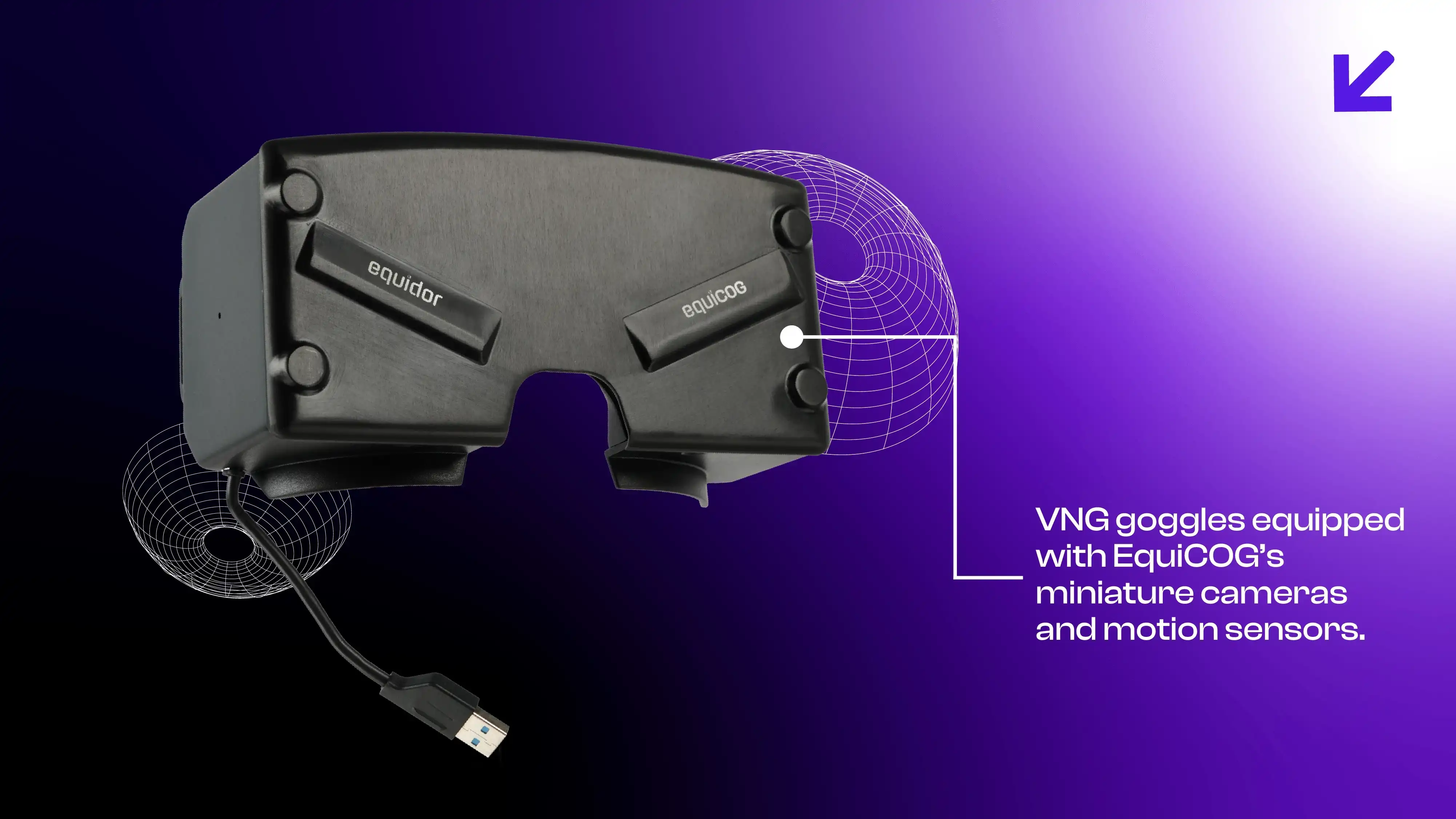
Advanced vestibular evaluation platforms such as EquiCOG combine high-resolution eye and head-tracking during comprehensive vestibular assessment, empowering ENT specialists to diagnose BPPV with unparalleled accuracy.
What Is BPPV?
BPPV is an inner-ear disorder where tiny calcium crystals dislodge and trigger brief episodes of spinning vertigo when the head changes position.2 Think of it as “grit in the gearbox” of your balance system: small particles that should be stationary start interfering with normal signals.
Symptoms & Signs
- Brief, intense spinning sensation when looking up, rolling over, or sitting up
- Nausea or vomiting
- Unsteady gait or imbalance
- Horizontal or torsional nystagmus on positional testing
Risks of Not Detecting and Treating
Untreated BPPV can lead to:
- Persistent falls and injuries
- Chronic imbalance and anxiety
- Reduced quality of life, driving avoidance, social withdrawal
Current Diagnostic Landscape
Standard vestibular evaluation often begins with a clinical history and the Dix–Hallpike manoeuvre.3 Many clinics rely on a simple VNG test using VNG goggles to visualize nystagmus. While helpful, traditional videonystagmography can miss subtle head-eye coordination deficits and torsional components, leading to incomplete vestibular assessment.
What’s Changing: The EquiCOG Innovation
EquiCOG elevates BPPV diagnosis through:
- Enhanced videonystagmography with simultaneous head tracking.
- 120 FPS high-frequency eye recording (captures every micro-nystagmus).
- Torsional eye movement tracing for intricate diagnostics.
- Real-time feedback and interactive prompts during positional tests.
- Configurable positional protocols that adapt to each patient.
Together, these features deliver a holistic vestibular evaluation, ensuring ENT doctors detect even the faintest positional nystagmus, critical for accurate BPPV diagnosis.
How EquiCOG Identifies BPPV
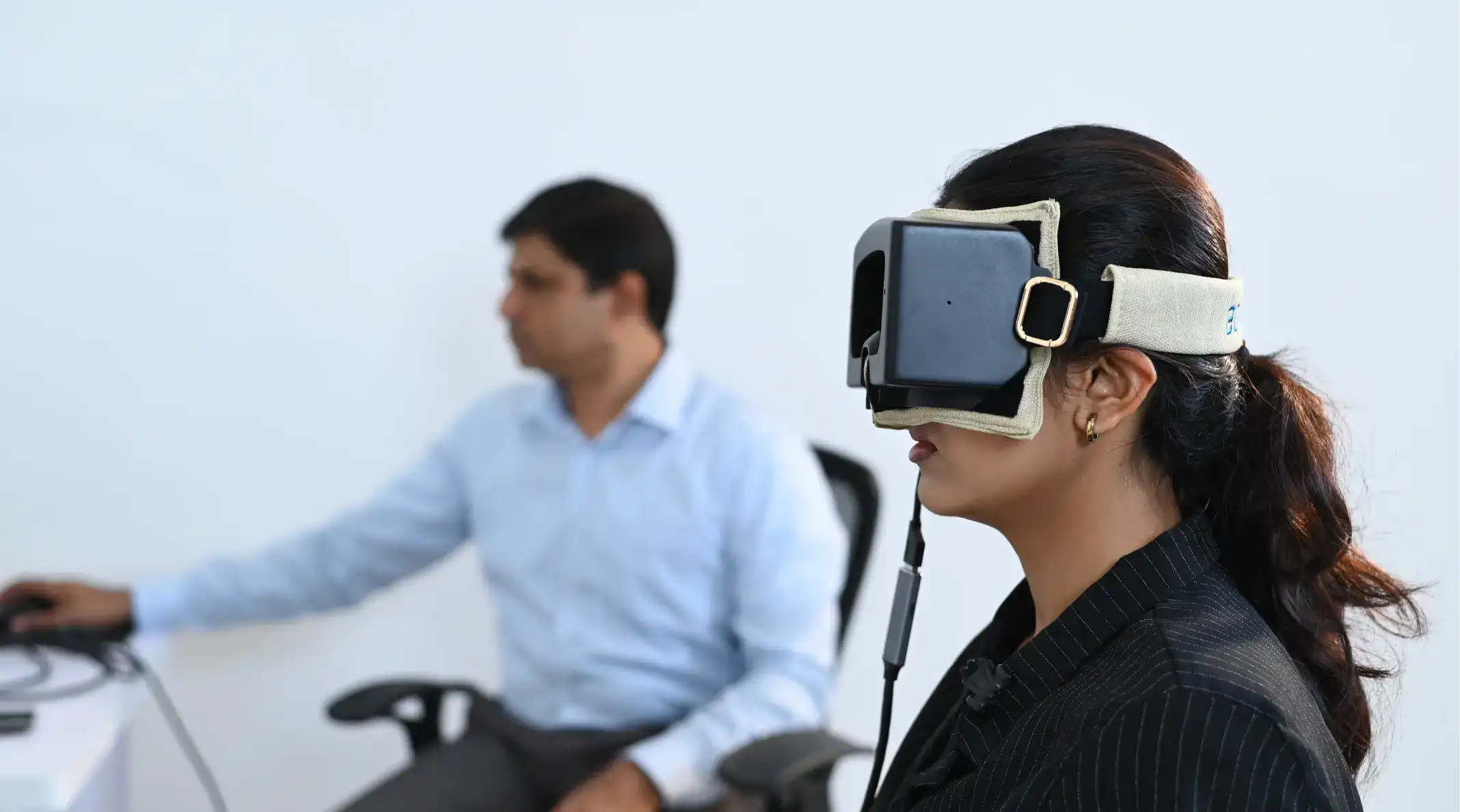
- The Patient wears lightweight VNG goggles equipped with EquiCOG’s miniature cameras and motion sensors.
- The clinician performs the Dix–Hallpike or Roll Test, guided by on-screen step prompts.
- EquiCOG’s dual tracking captures both head-position angles and eye movements in real time.
- Data is displayed as rectangular grid traces, velocity charts, and cumulative slow-phase velocities, highlighting the characteristic torsional nystagmus of posterior canal BPPV.
- ENT doctors review these precise metrics to confirm BPPV subtype and plan immediate canalith repositioning manoeuvres.
Call to Action
If dizziness is affecting you or your patients, insist on a comprehensive vestibular assessment with EquiCOG. Speak to your ENT specialist about advanced videonystagmography, or visit
<Taevas website> to download our brochure and request a demo. Early detection means quicker relief and a return to normal life.
References
- Swain, S. K., Munjal, S., & Shajahan, N. (2020). Vertigo in children: Our experiences at a tertiary care teaching hospital of eastern India. Journal of the Scientific Society, 47(2), 74-78.
- von Brevern M, Bertholon P, et al. Epidemiology of benign paroxysmal positional vertigo. J. Neurol. Neurosurg. Psychiatry. 2007.
- Bhattacharyya N, Baugh RF, et al. Clinical practice guideline: benign paroxysmal positional vertigo. Otolaryngol. Head Neck Surg. 2017.